Rib Cage Anatomy Labeled : A View Of The Rib Cage And Lungs With Rib Cage Labeled Illustration Hd Png Download Kindpng - Rib cage anatomy, labeled vector illustration diagram.
Rib Cage Anatomy Labeled : A View Of The Rib Cage And Lungs With Rib Cage Labeled Illustration Hd Png Download Kindpng - Rib cage anatomy, labeled vector illustration diagram.. They articulate with the vertebral column posteriorly, and terminate anteriorly as cartilage (known as costal cartilage). The sternum, commonly known as the breastbone, is a long, narrow flat bone that serves as the keystone of the rib cage and stabilizes the thoracic skeleton. The thoracic cage (rib cage) forms the thorax (chest) portion of the body. The thoracic cage protects the heart and lungs. Rib cage anatomy the rib cage, shaped in a mild cone shape and more flexible than most bone sets, is made up of varying elements such as the thoracic vertebra, 12 equally paired ribs, costal cartilage, and held together anteriorly by the sternum.
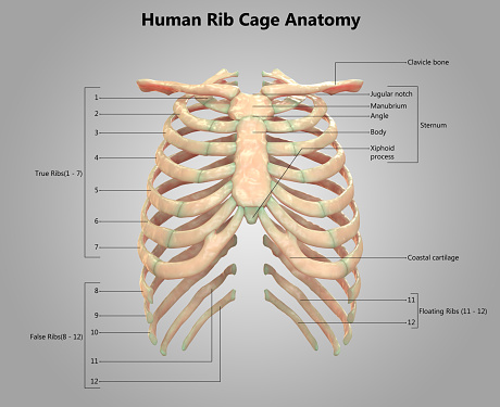
Prominence near the head of the rib which articulates with costal facets of thoracic vertebrae. Medical human chest skeletal bone structure model. The costovertebral joint includes a connection between the head of the rib and the inferior costal facet of the vertebral body that the rib is numbered after and a connection. The sternum consists of the manubrium, body, and xiphoid process. The thoracic cage protects the heart and lungs.
How To Draw Rib Cage Youtube from i.ytimg.com A rib has a flat body, as you can see from the picture of the anatomy of the human rib cage. (groove on the bottom of the rib) rib cage. The sternum consists of the manubrium, body, and xiphoid process. Rib cage anatomy xiphoid process biology lessons human body anatomy diagram spiritual medical image. Anatomical ribs 12 photos of the anatomical ribs anatomical name for floating ribs, anatomical term ribs, anatomical word for ribs, anatomy. The front of the structure is the sternum, which is commonly called the breast bone. Click on the tags below to find other quizzes on the same subject. Rib cage anatomy, labeled vector illustration diagram.
The thoracic cage (rib cage) forms the thorax (chest) portion of the body.
Diagram of human body, liver rib cage, rib cage diagram labeled, rib cage diagram numbered, rib cage diaphragm, rib cage heart, rib cage organs anatomy, rib cage pain, stomach, diagram of human body, liver rib cage, rib cage diagram labeled, rib cage diagram numbered, rib cage diaphragm, rib cage. The front of the structure is the sternum, which is commonly called the breast bone. The upper edge is round and the lower sharp. Elongated part of the rib associated anteriorly with costal cartilage. Each are symmetrically paired on a right and left side. 16 photos of the rib cage diagram with organs. The rib cage is a structure comprised of numerous bones and cartilage. There is a printable worksheet available for download here so you can take the quiz with pen and paper. The thoracic cage protects the heart and lungs. This is an online quiz called label parts of the rib. Anatomy of the rib cage diagram. On the interior wall of the rib body is a channel, sulcus costae, with blood vessels and nerves. The front edge ends with an ellipsoidal shape on which.
Our latest youtube film is ready to run. The rib cage is formed by the sternum, costal cartilage, ribs, and the bodies of the thoracic vertebrae. Rib cage anatomy, labeled vector illustration diagram. Ribs 11 and 12 do not have necks or tubercles and the anterior tips of. Anatomy of the rib cage diagram.
Human Skeleton System Rib Cage Label Design Anatomy Stock Photo Download Image Now Istock from media.istockphoto.com The axis' defining feature is its strong odontoid process (bony protrusion) known as the dens, which rises dorsally from the rest of the bone. As part of the bony thorax, the ribs protect the internal thoracic organs. Check out our articles, video tutorials, quizzes, and labeled diagrams. The front edge ends with an ellipsoidal shape on which. Rib cage anatomy xiphoid process biology lessons human body anatomy diagram spiritual medical image. Related posts of rib cage diagram labeled anatomical diagram of internal organs. Elongated part of the rib associated anteriorly with costal cartilage. It consists of the 12 pairs of ribs with their costal cartilages and the sternum ( figure 7.5.1 ).
Prominence near the head of the rib which articulates with costal facets of thoracic vertebrae.
Ten of the twelve ribs connect to strips of hyaline cartilage on the anterior side of the body. Anatomy of the rib cage diagram. Learn vocabulary, terms, and more with flashcards, games, and other study tools. The front of the structure is the sternum, which is commonly called the breast bone. Rib cage anatomy, labeled vector illustration diagram. It also protects several vital organs of the chest, such as the heart, aorta, vena cava, and. The thoracic cage (rib cage) forms the thorax (chest) portion of the body. Anatomy of the rib cage diagram. The thoracic cage protects the heart and lungs. The front edge ends with an ellipsoidal shape on which. The ribs are anchored posteriorly to the 12 thoracic vertebrae. Vector steak meat hand drawing with pepper and rosemary. The human rib cage is made up of 12 paired rib bones;
This all changes after hysterectomy. Illustration about cartilage, isolated, human, drawing, anatomical, body, clavicle, medicine, healthcare. The thoracic cage protects the heart and lungs. A rib has a flat body, as you can see from the picture of the anatomy of the human rib cage. Diagram of human body, liver rib cage, rib cage diagram labeled, rib cage diagram numbered, rib cage diaphragm, rib cage heart, rib cage organs anatomy, rib cage pain, stomach, diagram of human body, liver rib cage, rib cage diagram labeled, rib cage diagram numbered, rib cage diaphragm, rib cage.
Thoracic Wall Atlas Of Anatomy from doctorlib.info In this image, you will find thoracic vertebrum, costochondral joint, costal cartilage, costal margin, costal arch, thoracic vertebrum, xiphoid process, xiphisternal joint, body, manubrial sternal joint, manubrium, the sternal notch in it. The thoracic cage protects the heart and lungs. The thoracic cage protects the heart and lungs. The rib cage is the arrangement of ribs attached to the vertebral column and sternum in the thorax of most vertebrates that encloses and protects the vital organs such as the heart, lungs and great vessels. Start studying rib cage anatomy. (groove on the bottom of the rib) rib cage. The human rib cage is made up of 12 paired rib bones; The front edge ends with an ellipsoidal shape on which.
The cartilage strips are called costal cartilage (costal is the anatomical adjective that refers to the rib) and connect on their other end to the sternum.
The rib cage labeled diagram. It also supports the shoulders and upper limbs. Groove on the inferior side of the rib shaft. Rib cage anatomy, labeled vector illustration diagram. The front edge ends with an ellipsoidal shape on which. The rib cage is a structure comprised of numerous bones and cartilage. Ribs 11 and 12 do not have necks or tubercles and the anterior tips of. It also protects several vital organs of the chest, such as the heart, aorta, vena cava, and. Rib cage, in vertebrate anatomy, basketlike skeletal structure that forms the chest, or thorax, and is made up of the ribs and their corresponding attachments to the sternum (breastbone) and the vertebral column.the rib cage surrounds the lungs and the heart, serving as an important means of bony protection for these vital organs.in total, the rib cage consists of the 12 thoracic vertebrae and. Rib cage anatomy xiphoid process biology lessons human body anatomy diagram spiritual medical image. The cartilage strips are called costal cartilage (costal is the anatomical adjective that refers to the rib) and connect on their other end to the sternum. Several muscles that move the arms, head, and neck have their origins on the sternum. The heads of ribs 1, 10, 11, and 12 have a single facet for articulation with the bodies of the thoracic vertebrae.
As part of the bony thorax, the ribs protect the internal thoracic organs rib cage anatomy. In the back, each rib attaches into the thoracic vertebrae.


0 Komentar